The eyes
The eye is the organ that lets you see. Your eye is more than just the eyeball. Each eye is made up of the:
- eyeball (also called the globe)
- adnexal structures (also called accessory structures)
- eye socket
Most of the eye cannot be seen because it is covered by the surrounding adnexal structures and the bones of the skull that are part of the eye socket (orbit).
Each eye connects to the
The eyeball
The eyeball is also called the globe. It is a sphere about 2.4 cm (almost 1 inch) in diameter. The eyeball is made up of layers of tissue (the wall) that surround the lens and 2 chambers that support the eye and help maintain its shape. Each chamber is filled with a different type of fluid.
The aqueous humour is a clear, watery fluid that fills the space behind the cornea, in front of the lens (called the anterior chamber).
The vitreous humour is a clear, jelly-like fluid that fills the space behind the lens, in front of the retina (called the posterior chamber).
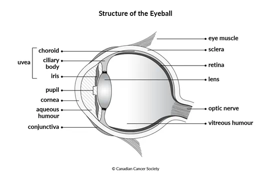
The wall of the eye
The wall of the eye is the outer part of the eyeball that surrounds the fluid-filled chambers inside the eye. It is made up of 3 layers (called tunics).
- The sclera and cornea make up the outermost layer.
- The uvea is the middle layer.
- The retina is the innermost layer.
Sclera
The sclera is the white part of the eye. It covers most of the outside of the eyeball. The sclera is made up of tough connective tissue and acts as a protective coating for the eye.
Cornea
The cornea is the clear, dome-shaped part at the front of the eye. It covers the pupil (the black, circular opening in the centre of the eye) and iris.
The cornea helps to direct and bend (refract) the light coming into the eye before it reaches the lens. The lens and cornea work together to allow you to see.
Uvea
The uvea is made up of 3 parts – the iris, the choroid and the ciliary body.
The uvea has many blood and lymph vessels that help bring nutrients to the eye. The muscles in the uvea help to control the amount of light coming into the eye before it reaches the lens and the direction of the light before it reaches the retina.
Iris
The iris is the coloured part of the eye that surrounds the pupil. The iris is made of connective tissue and muscle. The muscles of the iris change the size of the pupil to control the amount of light that enters the eye.
The colour of your iris comes from the amount of
Choroid
The choroid is a thin layer of tissue that lies between the sclera and retina. It contains many tiny blood vessels that bring oxygen and nutrients to the retina. The choroid also contains melanin that absorbs extra light entering the eye. This prevents reflections inside the eye that can impair vision.
Ciliary body
The ciliary body is a ring of muscle that extends out from the choroid at the point where the choroid meets the retina. It helps to hold the lens in place. The ciliary body is divided into 2 parts – the ciliary muscle and the ciliary process.
The ciliary muscle is responsible for changing the shape of the lens so you can focus on objects near or far.
The ciliary process is responsible for making and secreting aqueous humour.
Retina
The retina is the innermost part of the wall of the eye. It’s made up of many layers of nervous system cells, including neurons and light-sensitive cells called photoreceptors. There are 2 types of photoreceptors – rods and cones.
When you look at something, light enters the eye, focuses on the retina and activates the rods and cones. They send signals to the layers of neurons below them, which pass the signals along the optic nerve to your brain. Your brain then uses the signals to create an image of what you are looking at.
Lens
The lens is a transparent disc-shaped structure inside the eye. It is held in place behind the iris by the ciliary body. The shape of the lens changes to allow the eye to focus on objects near and far. When light passes through the lens, it is refracted and focused on the retina to create an image of what you are looking at.
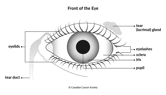
Adnexal (accessory) structures of the eye
The adnexal structures of the eye include the eyelids, conjunctiva and tear (lacrimal) gland. These structures protect, lubricate and support the eyeball. The adnexal structures of the eye are also called accessory structures of the eye.
Eyelids
The eyelids are folds of skin above and below each eye. They cover and protect the eye. Muscles around each eye control how the eyelids open and close. The sweeping motion of the eyelid across the eyeball when it opens and closes helps to remove any particles that may have gotten into your eye. When you open and close your eyes, you also help keep your eyes moist because the motion spreads tears across the exposed parts of the eyeball. The eyelids also have glands that produce an oily layer on the surface of the eyeball to stop the layer of tears from evaporating and the eye drying.
Eyelashes grow out of the edge of the eyelids. These also help protect the eye because they can stop sweat, dust and other particles from getting in your eyes.
Conjunctiva
The conjunctiva is a clear
Tear (lacrimal) gland
The tear gland (also called the lacrimal gland) is at the upper, outer corner of the eye. It secretes tears. When you blink, tears are swept across the surface of the eye. Tears lubricate the conjunctiva covering the surface of the eye and inner eyelid. Tears also remove dust and other particles from the eye and help to prevent infection.
Tears drain from the eye through very small openings in the inner corners of the upper and lower eyelids. They then travel down through the tear ducts and out the nose.
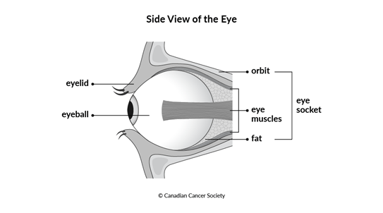
Eye socket
The orbit is the bowl-shaped structure in the skull where the eyeball sits. It also holds the surrounding muscles, nerves, fat, blood vessels, connective tissues and tear (lacrimal) gland.
The eye socket is made up of the orbit and the muscle and fat around the eyeball. The bone and other tissues of the eye socket help to cushion and protect the eyeball.
The eye muscles in the eye socket attach to the bones of the orbit on one end and to the sclera at the other. These muscles allow you to look around by controlling the movement of your eyeball.
Function of the eye
The eye and the brain work together to allow you to see. How they do this is very complex and depends on many parts and processes. The eye collects information about what you’re looking at in the form of light. The information that the light tells the rods and cones is then converted to a form of information that our brain can use. These signals are passed from the neurons in the retina to the optic nerve, which carries them to the brain. Your brain then processes the information in these signals and creates the image that you see.
Your eyes work together to allow you to see in 3D (called binocular vision). Binocular vision gives us the ability to determine depth and speed. If you lose the vision in one eye because the eyeball is removed or because a part of the eye is severely damaged, the other eye allows you to see most of what you could see before (though this depends on the vision in your remaining eye). But your ability to judge depth and speed will be affected.
Your trusted source for accurate cancer information
With support from readers like you, we can continue to provide the highest quality cancer information for over 100 types of cancer.
We’re here to ensure easy access to accurate cancer information for you and the millions of people who visit this website every year. But we can’t do it alone.
Every donation helps fund reliable cancer information, compassionate support services and the most promising research. Please give today because every contribution counts. Thank you.